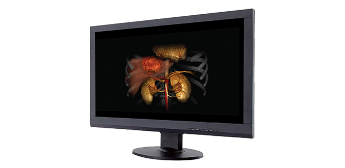
Image Guided Treatment Delivery
Image Guided Treatment Delivery | Interventional Oncology
Our sophisticated interventional oncology solutions can help you prioritize smart, efficient imaging, while optimizing safety and precision for every procedure.
One room. One system.
Many possibilities.
With a single, streamlined workflow, there’s no longer a need to transfer patients between rooms. Instead, you can now switch seamlessly between CT and angiography for a faster, safer, more comfortable process in one setting.

Alphenix solutions for interventional oncology
Alphenix floor and ceiling-mounted C-arms have been designed to support complex interventional procedures with unprecedented patient access and full-body coverage. Now, with our comprehensive range of planning, imaging chain, and dose management tools, you’ll have everything you need to plan, analyze and deliver the life-enhancing care your patients need.*
*Available on Alphenix workstation and Vitrea

Tumor Assessment with Whole Organ Perfusion
Volume Perfusion Liver*
With the ability to evaluate tumor response or anti-angiogenesis, CT body perfusion has become a valuable clinical tool – especially now that you’ve got 16 cm of z-axis coverage at your disposal.
Liver Perfusion Protocol: DLP = 720.8 mGy.cm, Effective dose = 10.81 mSv (k = 0.015)
*CT Volume perfusion only available with Aquilion ONE

Tumor Assessment Applications
Transform your workflow with these innovative tools:
Learn more about CT Clinical Applications >
Learn more about Angiography Clinical Applications >
Learn more about Multi-modality Clinical Applications >
* CT Volume perfusion only available with Aquilion ONE

CME Webinar
Image Guided Interventional Radiology Procedures Using a Combined Angiography CT System
David Hays, MD & Farah Gillen Irani, MD

CME Webinar
Improving Oncologic Outcomes Using a Unique 4DCT Solution
Junsung Choi, MD
Image Guided Treatment Delivery | CT Fluoroscopy
Interventional CT
Simple and streamlined CT Fluoroscopy
Conduct faster, more focused interventional procedures with a new hybrid CT Fluoroscopy interface that enables one-handed operation with ergonomically designed controls and a versatile touchscreen tablet.

Image Guided Treatment Delivery | Ultrasound
Dedicated transducers and an abundance of imaging and navigation tools help you enhance clinical confidence and accuracy during interventional procedures and their follow-up.
Aplio i-series
Clear Image for Precise Procedures
Canon Medical Systems’ flagship ultrasound systems, the Aplio i-series, is designed to deliver outstanding clinical precision with incredible staff productivity. Crystal-clear images, with enhanced resolution and penetration also help you perform accurate biopsies for more detailed treatment delivery guidance.

Enhanced Visualization During Biopsies
Biopsy Enhancement Auto Mode (BEAM)
One-button needle enhancement helps minimize complications while increasing visual accuracy during interventional procedures.
- Standard on most linear transducers
- No sacrifice of image quality

Smart Navigation
We’ve created a range of technologies that can help improve your clinical confidence during ultrasound-guided interventional procedures.
- Displays virtual biopsy lines that correspond with needle positions on the fused live image
- Improves visualization of the needle tip for up to three biopsy needles
- Can be used in conjunction with CIVCO’s virtuTRAX for Aplio 500 Platinum and omniTRAX for i-series

Simplify Your Workflow
Smart Fusion Technology*
Smart Fusion* offers the best of both worlds by synchronizing ultrasound with CT or MRI images to help you locate hard-to-find lesions. It works by reading CT/MRI/US DICOM data sets using position sensors located on the ultrasound transducer to display the corresponding images side-by-side. This can help you boost efficiency and minimize dose during interventional procedures.
*Available on Aplio 500 Platinum and i-series


Merging modalities to improve confidence
Matching the transducer position with the pre-acquired 3D data set is a simple and quick two-step process. By moving the transducer over the region of interest you can now browse the area simultaneously in both real-time ultrasound and pre-acquired volume data. Intelligent target and marker points facilitate navigation in the region of interest.
Prostate Fusion Biopsy

Fusion Ovary


